Description about this room/facility
Location
Located in the Medical Sciences Building- H519
Rutgers University

Pinnacle 8400 Rat Tethered System
The Pinnacle 8400 rat tethered system it provides information about electroencephalographic readings that can shed light on brain oscillations network excitability and seizure information (EEG Set-up)
The Pinnacle 8400 rat tethered system located in MSB H519 allows for a configurable channel setup and is currently capable of recording simultaneously from 2 animals. It can obtain 4 EEG signals as well as a single depth recording of seizure activity. The EEG data are amplified and filtered by a headmounted 10x preamplifier, which greatly reduces electrical noise. Signals are then passed through the rat commutator/swivel to the final stage conditioning and filtering via a data acquisition system, ADInstruments Powerlab 16/35. The data are then transmitted real-time to a data recording, signal processing software, Labchart. In conjunction with the EEG data, video recordings are recorded with via a high resolution infrared dome camera which can record an object up to 65ft. away in the day or nighttime and synchronized with the EEG data in Labchart. The recorded movie and data file can be played back to view the recorded video and EEG data file simultaneously. Signal analysis and filtering are all performed in LabChart.
The Pinnacle 8400 rat tethered system located in MSB H519 allows for a configurable channel setup and is currently capable of recording simultaneously from 2 animals. It can obtain 4 EEG signals as well as a single depth recording of seizure activity. The EEG data are amplified and filtered by a headmounted 10x preamplifier, which greatly reduces electrical noise. Signals are then passed through the rat commutator/swivel to the final stage conditioning and filtering via a data acquisition system, ADInstruments Powerlab 16/35. The data are then transmitted real-time to a data recording, signal processing software, Labchart. In conjunction with the EEG data, video recordings are recorded with via a high resolution infrared dome camera which can record an object up to 65ft. away in the day or nighttime and synchronized with the EEG data in Labchart. The recorded movie and data file can be played back to view the recorded video and EEG data file simultaneously. Signal analysis and filtering are all performed in LabChart.

Whole Cell Path Configuration
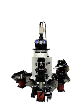
Located in the MSB H519 (Rutgers University), the experimental system resting on a TMC vibration isolation table consists of an Olympus BX41 upright microscope designed specifically for the rigorous demands of challenging electrophysiological experiments. The microscope has infrared/DIC optics, epifluorescence illumination with a turret designed for 6 cubes and is coupled to 1/2" Interline CCD camera (380-1200 NM, 570 lines) and a 1000-line, 2-channel video monitor. System runs NIS Elements for image acquisition and control of illumination sequences. The microscope is mounted on a Sutter Instruments motorized stage translator.
A Scientifica PatchStar ultra stable, super smooth micromanipulators with X, Y, Z and a smart-sensor- based virtual approach axes is mounted on a Tholabs gantry system. The platform supports a PM-1 Submerged Slice Chamber (Warner Instruments) with perfusion flow is accomplished with a low-electrical noise, two channel peristaltic pump (Watson-Marlow, 400 series) coupled to a 1 channel feed-back temperature control system (TC-324B, Warner Instruments) and Inline Solution Heater (SH-27B, Warne Instruments) for maintaining recorded slices at physiological temperatures.
Recording are obtained using an Axon Multiclamp 700B amplifier (Molecular Devices), a computer- controlled, dual channel, resistive-feedback patch clamp and high-speed current clamp with two CV- 7B low-noise headstages. The amplifier is coupled to a Axon Digidata 1400A (Molecular Devices), a low-noise high-resolution 16-bit data acquisition system with a maximum sampling rate of 250 kHz per channel for electrophysiology recordings connected to an USB 2.0 interface on a Dell Optiplex 780 desktop running pCLAMP 10 (Molecular Devices) Data Acquisition and Analysis Software capable of automated execution of protocols, recording in gap-free and episodic waveform stimulation, online filtering and leak subtraction and online statistical analysis. A 6L Circulating Water-bath (Cole-Parmer) is used for pre heating solutions which are connected to cylinders for constant source of oxygen. Additionally, the system includes a High Voltage, Bipolar Rechargeable Stimulus Isolator (WPI A365R) designed for stimulus delivery in neurophysiological applications and a 2 channel Tektronix oscilloscope.