Need a description of this room/ facility
Location
Need a location for this room/facility

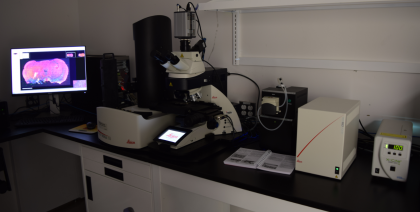
Leica Aperio Versa 200: Brightfield, Fluorescence & FISH Digital Pathology Scanner
Purpose: To digitally scan mounted tissue samples from 1.25X to 63X magnification. Equipped with a 200-slide autoloader and a comprehensive range of image analysis solutions, the slide scanner will be used to autonomously scan holistic brain tissue, saving hours of user viewing and analysis time.
Capabilities: This microscope system is capable of viewing samples at 1.25X, 5X, 40X, and 63X magnification for both brightfield and fluorescence imaging.
Coronal brain slice of injured rat model. Blue stain represents cell nuclei (DAPI), green stain represents neurons (NeuN), and red stain represents area of extracellular matrix degradation (MMP2).
Unique Features:
200 slide autoloader
1.5X, 5X, 40X, 63X magnification
Automated oiler and oil-immersion lenses
One-click scan protocols
7 filter positions

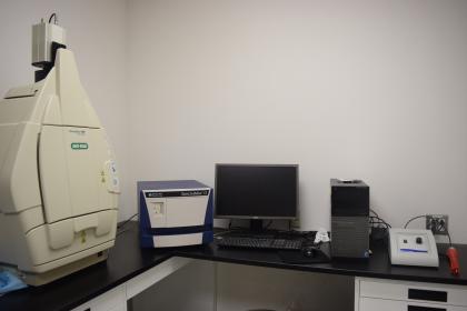
ChemiDoc™ MP System
The ChemiDoc MP system is a full-feature instrument for gel or western blot imaging. It is designed to address multiplex fluorescent western blotting, chemiluminescence detection, and general gel documentation applications.
Its features are based on CCD high-resolution, high-sensitivity detection technology, and modular options to accommodate a wide range of samples and support multiple detection methods. The system is controlled by Image Lab™ software to optimize performance for fast, integrated, and automated image capture and analysis of various samples.
Features and Benefits
Multiple imaging capabilities — the ChemiDoc MP imager can accommodate a variety of sample types and detection methods including multiplex fluorescent western blotting. It is the perfect imager to accompany your protein and DNA electrophoresis runs as well as your western blotting experiments. It delivers quantitative, reproducible results for fluorescence, chemiluminescence, and colorimetric detection
Stain-free technology — UV-induced fluorescence labeling of proteins in the stain-free gels allows a 2 hr Coomassie gel-staining protocol to be condensed into a 5 min stain-and-image step. Stain-free gels are western blot compatible — using the V3 Western Workflow™, check your electrophoresis results and blot transfer quality prior to western blotting
High-sensitivity blot detection — the ChemiDoc MP imaging system offers advanced detection technology that determines optimal exposure, even for faint or intense samples. Superior sensitivity is achieved for chemiluminescence and multiplex fluorescence detection and for colorimetric gel and blot documentation
Superior Image Quality — exceptional dynamic range enables visualization of faint and intense bands on same blot or gel. Images are always in focus at any zoom level to ensure publication-ready images in seconds
Ease of Use — precalibrated system provides the precise focus for any zoom setting or sample height; automated hands-free operation ensures consistent, reproducible, and high-throughput performance

SpectraMax i3
The purpose of the SpectraMax i3 is to provide rapid data collection of microplate readings, using the SoftMax Pro Software
Capabilities:
This device has the capability of detecting: luminescence, absorbance, and fluorescence. The system allows the user to read 6-384 well microplates simultaneously.
The SpectraMax i3 is specifically used in this lab for BCA assays and Elisa Assays. It allows the user to determine the total concentration of protein in a solution. These concentrations are then used to perform western blots. The SpectraMax i3 also allows the user to identify antibodies during Elisa Assays.
The SoftMax Pro Software quickly captures and analyzes results from the SpectraMax i3, through different data analysis options. The software has ready to run protocols to read the microplates, different curve fit options, and provides results that are ready to be used.

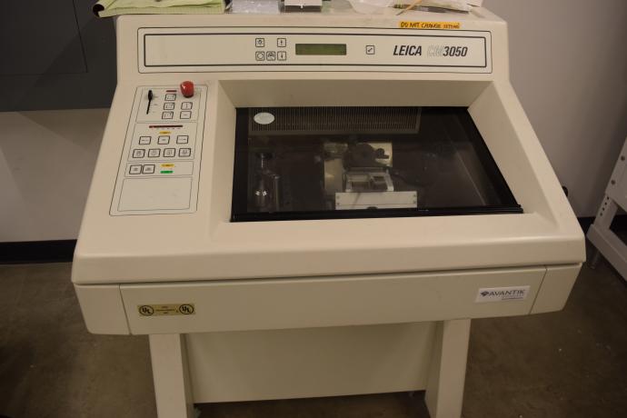
Leica CM3050 Research Cryostat
The Research Cryostat is primarily designed for cyrosectioning of delicate samples such as brain samples. The apparatus maintains a very low temperature making fine sectioning easier to do.